


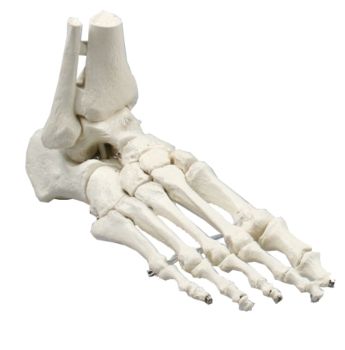
Bones of the Foot Anatomical Model
Articulated anatomical life size model. Educational Tool for students and clinics.

Product Description
Life-size Bones of the Foot Anatomical Model
A detailed, articulated representation of the adult foot skeleton designed to support clear anatomical understanding in educational and clinical environments.
Best for: anatomy education, podiatry clinics, patient consultations, and classroom demonstration.
Not ideal for: decorative display only, or users seeking a non-anatomical novelty model.
- Life-size proportions based on an adult foot
- Articulated joints for realistic movement
- Supplied with a detachable display stand
What is it?
The Bones of the Foot Anatomical Model is a life-size, articulated skeletal model of the adult human foot. It presents the individual bones in their correct anatomical relationships, allowing movement that reflects natural joint articulation.
The model includes the lower ends of the tibia and fibula, providing important context for the ankle joint and its connection to the foot.
Manufactured as a natural casting of an adult mobile foot, this model is intended to closely represent real anatomical structure rather than a simplified or abstract form.
Each bone is clearly defined, making it suitable for both teaching and demonstration purposes. The articulation allows users to observe how bones interact during movement, supporting a deeper understanding of foot mechanics.
A metal pin system allows the model to be mounted securely on the included stand or removed easily for handheld use. This flexibility makes it suitable for a range of settings, from desks and shelves to treatment rooms and classrooms.
Who is it for?
This anatomical model is designed for a broad audience that includes both healthcare professionals and learners. It is particularly useful for those who need a clear, tangible reference to explain or study foot structure.
- Podiatry and foot health practitioners explaining conditions or treatments
- Physiotherapists and other allied health professionals
- Students studying anatomy, podiatry, or related healthcare subjects
- Educators teaching musculoskeletal or lower limb anatomy
- Clinics that want a visual aid to support patient communication
It can also be helpful for informed patients who want to better understand their own foot anatomy when discussing care options with a professional.
What does it help with?
The model supports learning and communication rather than treatment. By providing a physical reference, it can make complex anatomical concepts easier to grasp.
- Understanding foot structure: Helps users visualise how many bones make up the foot and how they are arranged.
- Ankle–foot relationship: Shows how the tibia and fibula connect to the foot at the ankle.
- Joint movement: Articulation allows demonstration of how bones move relative to one another.
- Patient education: Can make explanations clearer during consultations.
- Study and revision: Provides a hands-on learning tool for students.
How does it work?
The model works by accurately replicating the bones of the foot in a durable, three-dimensional form. Articulated joints allow controlled movement, helping users observe motion pathways without the distraction of soft tissue.
The inclusion of the tibia and fibula provides anatomical continuity from the lower leg into the foot.
The detachable stand supports stable display at a comfortable viewing height. When removed from the stand, the model can be handled directly, allowing closer inspection of individual bones and joints. This dual-use design makes it adaptable to different teaching and demonstration styles
Problem → solution overview
| Common problem | How the model helps |
|---|---|
| Difficulty visualising foot bone structure | Provides a clear, three-dimensional view of individual bones |
| Explaining anatomy verbally takes too long | Offers an immediate visual reference during explanations |
| Flat diagrams lack depth and movement | Articulated joints show how bones move together |
| Patients struggle to understand the ankle–foot connection | Includes tibia and fibula insertion for anatomical context |
Key benefits
- Life-size scale supports realistic learning
- Articulated design allows demonstration of movement
- Detachable stand offers flexible display options
- Clear bone definition aids study and explanation
- Suitable for both professional and educational settings
- Supplied with storage box to keep components together
When using the model, ensure it is positioned securely on the stand or held firmly when removed. The model is designed to be handled gently; articulation should never require force. A stable setup supports clearer demonstrations and reduces the risk of accidental damage.
Common mistakes to avoid
- Forcing joints beyond their natural range of movement
- Assuming the model represents soft tissue or muscle detail
- Using the model as a weight-bearing or load-testing object
- Storing the model loose without the protective box
How to use
- Remove the model and stand from the storage box.
- If using the stand, align the metal pin with the hole in the model and insert securely.
- Place the stand on a flat, stable surface.
- Gently articulate the foot to demonstrate specific joints or movements.
- For handheld use, remove the model from the stand and support it with both hands.
- After use, return the model and stand to the box for safe storage.
Technical specifications
| Product type | Anatomical skeletal foot model |
|---|---|
| Scale | Life size (adult) |
| Articulation | Yes, movable joints |
| Included bones | Foot bones with tibia and fibula insertion |
| Stand | Included, detachable via metal pin |
| Use orientation | Universal (left/right representation) |
| Materials | Durable anatomical casting material (specific composition varies) |
| Care and cleaning | Wipe clean with a dry or lightly damp cloth |
| Pack contents | Foot model, display stand, storage box |
| Warranty | 5 years |
Frequently asked questions
Is this model suitable for patient consultations?
Yes. The clear bone definition and life-size scale make it suitable for helping patients visualise foot anatomy during discussions.
Can the model be used without the stand?
Yes. The stand is detachable, allowing the model to be used handheld when preferred.
Does it show muscles or soft tissue?
No. This model focuses on the skeletal structure of the foot and ankle only.
Is the articulation adjustable?
The joints are articulated to allow natural movement, but they are not adjustable beyond their intended range.
Is it appropriate for classroom use?
Yes. It is suitable for individual study or group demonstration in educational settings.
How durable is the model?
It is designed for repeated educational use when handled with care and stored properly.
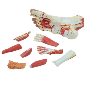
Complementary / pairing suggestions
- Ankle joint anatomical models for broader lower limb context
- Flat foot or high arch demonstration models
- Educational charts showing foot bone names and landmarks
Frequently bought together

Sold: Various Pack Sizes
Incl. VAT Exl. VAT
Sold: Each
Incl. VAT Exl. VAT
Sold: Each
Incl. VAT Exl. VAT
Sold: Each
Incl. VAT Exl. VAT
Sold: Each
Incl. VAT Exl. VAT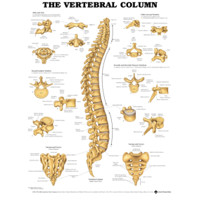
Sold: Each
Incl. VAT Exl. VATSold: Each
Incl. VAT Exl. VAT
Sold: Each
Incl. VAT Exl. VAT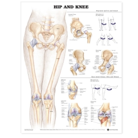
Sold: Each
Incl. VAT Exl. VATSold: Each
Incl. VAT Exl. VAT
Sold: Each
Incl. VAT Exl. VATSold: Each
Incl. VAT Exl. VAT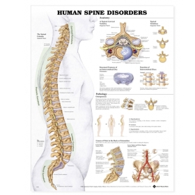
Sold: Each
Incl. VAT Exl. VAT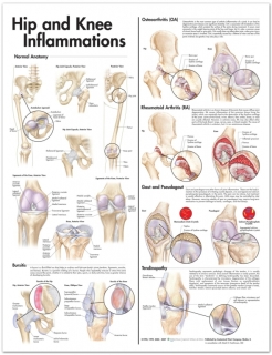
Sold: Each
Incl. VAT Exl. VAT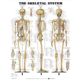
Sold: Each
Incl. VAT Exl. VAT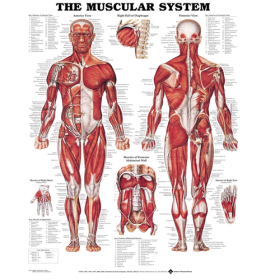
Sold: Each
Incl. VAT Exl. VATSold: Each
Incl. VAT Exl. VAT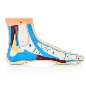
Sold: Each
Incl. VAT Exl. VAT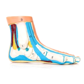
Sold: Each
Incl. VAT Exl. VAT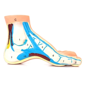
Sold: Each
Incl. VAT Exl. VAT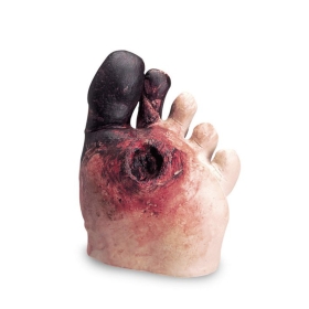
Sold: Each
Incl. VAT Exl. VAT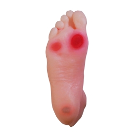
Sold: Each
Incl. VAT Exl. VAT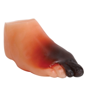
Sold: Each
Incl. VAT Exl. VAT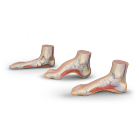
Sold: Set of 3
Incl. VAT Exl. VAT
Sold: Each
Incl. VAT Exl. VAT
Sold: Each
Incl. VAT Exl. VAT
Sold: Each
Incl. VAT Exl. VAT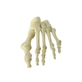
Sold: Each
Incl. VAT Exl. VAT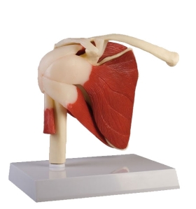
Sold: Each
Incl. VAT Exl. VAT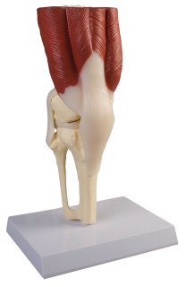
Sold: Each
Incl. VAT Exl. VAT
Sold: Each
Incl. VAT Exl. VAT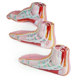
Sold: Set
Incl. VAT Exl. VATSold: Set
Incl. VAT Exl. VAT
Sold: With stand
Incl. VAT Exl. VAT