


Dermatoscope MoleScope II
Our dermatoscope with high magnification and cross-polarized light delivers high-resolution imaging to diagnose abnormally looking skin and nails.
Professional dermatoscopy tool for podiatrists.

Product Description
Professional Podiatry Dermatoscopy
MoleScope II is a battery-powered dermatoscope that enables the Podiatrist to expand the visual diagnosis of abnormally looking skin and nails, adding value to routine and follow-up examinations, as well as preventing further complications.
The digital images can be saved and help detect malignant changes over time. The device polarises reflected light to allow the examiner to see through the stratum corneum, visualising the deep layers of the epidermis and papillary dermis.
This allows examination of the characteristic pigment and vascular patterns of the skin as well as the architecture of the upper dermal papillae and rete ridges of the skin and nail bed (1).
Best for: Podiatrists seeking enhanced visual assessment, documentation, and monitoring of skin and nail conditions during routine and follow-up examinations.
Not ideal for: Situations requiring standalone imaging without a compatible smartphone or where advanced laboratory diagnostics are required.
- Designed for non-invasive clinical assessment
- Compatible with a wide range of modern smartphones
- Supports visual documentation over time
What is it?
MoleScope II is a battery-powered, handheld digital dermatoscope developed to extend the visual diagnostic capabilities of podiatrists and other foot health professionals. By combining high magnification with cross-polarised illumination, it enables close inspection of skin and nail structures that are not visible to the naked eye.
The device attaches to compatible smartphones and uses their camera systems to capture detailed images and videos. This approach allows clinicians to integrate dermatoscopy into everyday practice without the need for bulky imaging equipment or complex installation. The system is designed to be reusable and does not require direct skin contact when using the non-contact cap.
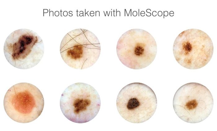
Cross-polarised light reduces surface reflection, allowing visualisation beneath the stratum corneum. This makes it possible to examine pigment distribution, vascular patterns, and structural features of the epidermis and superficial dermis, including nail beds.
Who is it for?
MoleScope II is intended for professional use in clinical, educational, and specialist environments where accurate visual assessment of foot-related skin and nail conditions is required.
- Podiatrists and podiatric surgeons
- NHS and hospital-based foot care services
- Private podiatry and multidisciplinary clinics
- Sports podiatrists and MSK practitioners
- Universities, teaching hospitals, and training providers
- Specialist clinics involved in skin and nail monitoring
What is it used for?
The MoleScope II is used as a clinical aid to examine a wide range of pigmented and non-pigmented skin and nail presentations commonly encountered in podiatry. It supports both initial assessment and ongoing monitoring by allowing consistent visual comparison over time.
Typical applications
- Assessment of pigmented lesions on the foot and ankle
- Evaluation of non-pigmented cutaneous conditions
- Examination of nail plate and nail bed abnormalities
- Monitoring of lesions during follow-up appointments
- Clinical documentation and patient records
Characteristics
- High magnification imaging suitable for detailed inspection
- Cross-polarised LED lighting to minimise surface glare
- Non-invasive and reusable design
- Portable and battery powered for clinic or outreach use
Typical users
- Clinicians performing routine foot skin assessments
- Podiatrists monitoring lesion changes over time
- Educators demonstrating dermatoscopic features
- Clinics aiming to enhance visual diagnostic capability
How to work with it
The device is attached to a compatible smartphone using the supplied universal attachment. Once aligned with the camera, the dermatoscope can be activated to illuminate and magnify the area of interest.
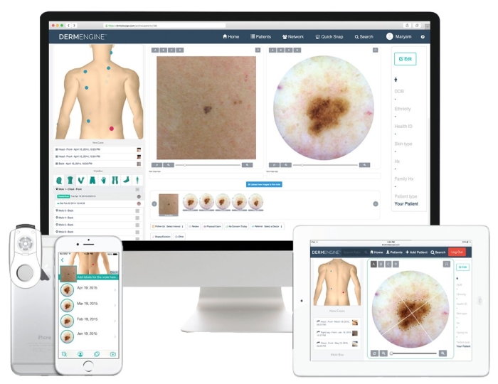
Images are captured using the accompanying software, enabling storage and review as part of the patient record.
Problem and solution overview
| Common problem | How the product helps |
|---|---|
| Limited visibility of subsurface skin structures | Cross-polarised light reduces reflection and allows deeper visual assessment |
| Difficulty documenting skin changes over time | Digital image capture supports visual records and follow-up comparison |
| Uncertainty when assessing pigmented lesions | Magnification highlights pigment patterns and vascular features |
| Need for non-invasive assessment tools | Reusable, non-contact imaging avoids skin trauma |
Benefits for Podiatrists
The study published in Journal of Foot and Ankle Research highlights the significant benefits of using a dermatoscope in podiatric practice, especially for diagnosing and managing various skin and nail conditions.
A dermatoscope provides podiatrists with an enhanced view of the skin’s surface, allowing for closer inspection of pigmentation, texture, and vascular structures. This tool is invaluable for identifying issues such as melanocytic lesions, fungal infections, and other abnormalities that may not be visible to the naked eye.
By magnifying and illuminating the skin, a dermatoscope helps podiatrists make more accurate, timely diagnoses, which is essential for early intervention and effective treatment planning.
Furthermore, the dermatoscope’s ability to visualise intricate skin and nail features enhances the podiatrist’s understanding of a patient’s condition, aiding in the differentiation between benign and potentially malignant lesions.
Podiatrist Monitoring Tool
For podiatrists, this level of detail can be particularly helpful in monitoring conditions like plantar warts, psoriasis, and other dermatological concerns affecting the feet.
As a diagnostic aid, it improves the precision of patient assessments and can reduce the need for invasive procedures. This ultimately supports better patient outcomes by fostering early intervention strategies, which are crucial for conditions that may worsen over time if left undetected.
Molescope ll Key benefits
- Enhances visual assessment beyond the naked eye
- Supports early identification of suspicious features
- Improves clinical documentation and monitoring
- Portable design suitable for varied clinical settings
- No subscription or recurring software costs
- Compatible with widely used smartphone platforms
Common mistakes to avoid
- Assuming dermatoscopy replaces clinical judgement or referral pathways
- Using poor image alignment, leading to unclear captures
- Over-interpreting single images without follow-up context
- Neglecting routine device charging and maintenance
How to work with MoleScope II
- Fully charge the device before initial use.
- Attach the dermatoscope securely to the smartphone using the universal mount.
- Launch the compatible imaging software.
- Position the device over the area of interest, using the appropriate cap.
- Activate the LED lighting and capture images as required.
- Store and label images according to clinic documentation protocols.
Technical specifications
| Magnification | Approximately 60× (varies by smartphone model) |
|---|---|
| Field of view | Approximately 16 mm |
| Lighting | 6 white LEDs with cross-polarisation |
| Polarisation coefficient | High contrast polariser (approx. 99.97%) |
| Battery | Rechargeable lithium-ion |
| Battery life | Up to 6 hours working time |
| Charging | USB 2.0 with charge and low battery indicator |
| Caps | Glass contact plate with scale and non-contact cap |
| Compatibility | Selected iPhone, iPad, iPod, and Samsung Galaxy models |
| Regulatory status | Medical Device Class I, UKCA and CE marked |
| Warranty | 1 year |
Frequently asked questions
Can MoleScope II diagnose skin cancer?
No. MoleScope II is a visual assessment and documentation tool. It may help identify features that warrant further investigation or referral but does not provide a diagnosis.
Is the device suitable for nail examination?
Yes. The magnification and lighting are suitable for examining nail plates and nail beds, including changes in colour, structure, and vascularity.
Does it require direct skin contact?
No. The device includes a non-contact cap, allowing imaging without touching the skin when appropriate.
Can images be stored for long-term monitoring?
Yes. Images can be saved using the accompanying software, supporting comparison over time during follow-up visits.
Is the software subscription-based?
No. The included software does not require subscriptions or additional purchases.
How durable is the device for clinical use?
The device is designed for repeated professional use when handled and maintained according to guidance.
Complementary and pairing suggestions
- Clinical photography documentation systems
- Podiatry examination lighting and magnification tools
- Patient record and imaging software solutions
- Disposable clinical hygiene accessories
Written by: Algeos Product & Clinical Content Team
Last reviewed: February 2026
Purpose: This content is provided for buyer guidance and product understanding only. It does not replace professional training, diagnosis, or medical advice.
Frequently bought together
Sold: Pack of 10
Incl. VAT Exl. VAT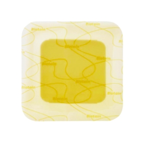
Sold: Various Pack Sizes
Incl. VAT Exl. VAT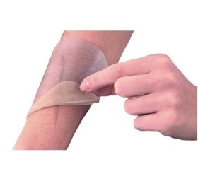
Sold: each
Incl. VAT Exl. VAT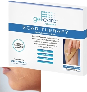
Sold: 1 or 10 Sheets
Incl. VAT Exl. VAT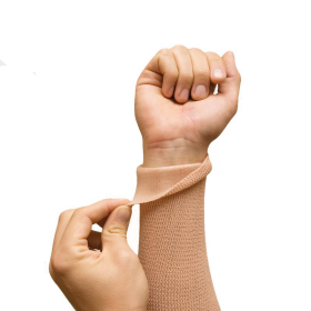
Sold: Each
Incl. VAT Exl. VAT
Sold: One roll
Incl. VAT Exl. VAT
Sold: Each
Incl. VAT Exl. VAT